Summary
A method for accurate, efficient and non-invasive assessment of the pressure drop in the cardiovascular system using time-varying blood velocity profiles from from any flow imaging techniques. This technology will improve the screening power of current clinical practice and will reduce the need for catheterisation procedures.
What
A technology that allows an accurate estimate of pressure drops within the cardiovascular system from blood flow imaging, also in the presence of blood flow constrictions. The technology has been designed to work with any imaging techniques able to measure 2D time-variable blood flow profiles along a given section, such as 2D Doppler echocardiography (ECG), 3D Doppler and 2D or 3D PC-MRI. The technology can be used for improving risk stratification and clinical decision-making in several conditions.
Why
Assessment of pressure variations in the cardiovascular system is critical for diagnosis and treatment planning of many cardiovascular diseases.
To date, catheterisation is the gold standard for measuring pressure drop in the cardiovascular system. However, catheterisation is an invasive technique associated with high risks of infection and high costs. As a non-invasive alternative, the simplified Bernoulli principle allows to estimate the pressure drop along a vessel as a function of a single peak velocity event. However, the overly simplified assumptions behind the Bernoulli principle do not allow to achieve a correct prediction in different cases, such as in stenotic vessels (i. e. in the presence of flow constrictions).
Other known alternative methods for estimating a blood pressure drop are based on computer simulations of the three-dimensional blood flow in vascular structures. However, the simulations require time, a skilled engineer and high computational resources, and their results are dependent on anatomical details and boundary conditions that are difficult to extract from images.
Hence, an accurate, cost-effective and non-invasive method for estimating pressure differences in blood vessels in real time has been a long-felt need.
Benefits
The proposed technology enables to accurately estimate the pressure drop not only in normal laminar flow, but also across a flow constriction, such as a stenosis. The data can be processed in real time during imaging acquisition, with no associated risk for the patient. The technology can be easily integrated in existing imaging scanning software and/or in advanced PACS software.
The technology has been specifically designed to work with any imaging technique, able to measure the entire velocity profile along at least a section of a vessel. This means that it can work with partial observations available through 2D Doppler ECG, as well as PC-MRI flow imaging techniques, or any complete acquisition of the flow made using recent developments of echocardiography (e.g. speckle decorrelation techniques).
Opportunity
The technology has been tested and validated in different body districts, including aortic valves and patients with hypoplastic left heart syndrome.
The technology is protected by a European patent application, a US granted patent, a US continuation patent application, and a Chinese patent application and is available for licensing. Suitable commercial partners are sought for integration and commercialisation of the technology.
The Science
An accurate estimate of a pressure drop along any normal or stenotic vessel can be derived considering only the advective components of the forces acting on the blood flow. This is conceptually a correction of the commonly used simplified Bernoulli equation by accounting for the complete velocity profile. Specifically, the advective components are calculated as a function of the whole velocity profile measured along one or more sections of interest.
For a more accurate result, the velocity field can be measured on an inlet section and on an outlet section, and the advective components of the velocity fields can be calculated along both the measured sections. However, the discovered function can also use only blood flow velocity profiles measured at a given section, and preferably at the section experiencing the maximum blood flow velocity (e.g. on constrictions), without the need of any further measurement on any other section. The estimated pressure drop can be used as a metric to evaluate the conduit function of a vessel at any point along its length.
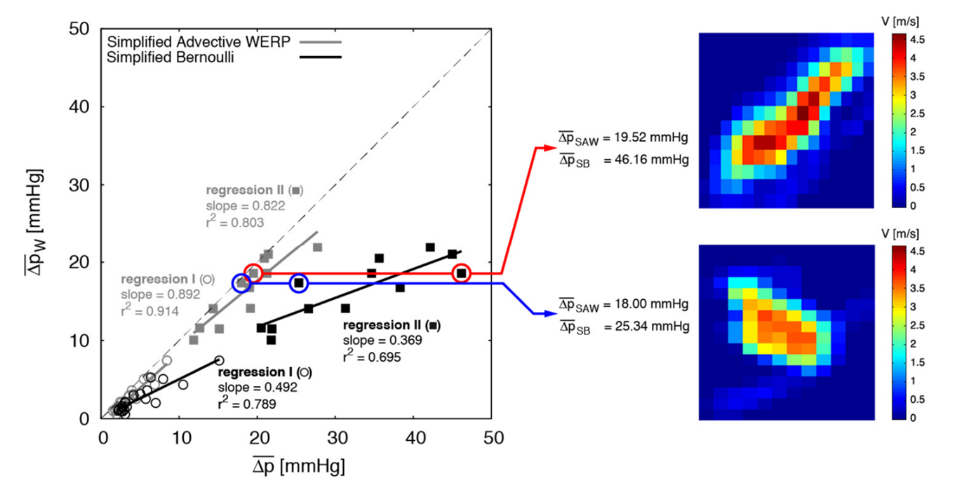
Figure: Comparison between Simplified Bernoulli (SB) and the proposed new method for calculating pressure drop with measurements taken only on a given outlet section (here referred as Simplified Advective WERP – SAW). Note how the estimate of the pressure drop using the SAW method leads to values very close to reality in the two selected cases (blue and red circles), whereas SB introduces a spurious difference between these two cases
IP Status
The technology is protected by:
Further Information
Donati, F. et.al., (2017), "Beyond Bernoulli: Improving the Accuracy and Precision of Noninvasive Estimation of Peak Pressure Drops", Circulation: Cardiovascular Imaging, 10(1), e005207, doi:10.1161/circimaging.116.005207.
De Vecchi, A. et.al., (2022), "Unlocking the Non-invasive Assessment of Conduit and Reservoir Function in the Aorta". Journal of Cardiovascular Translational Research, doi:10.1007/s12265-022-10221-4