Summary: A clinically validated tool to identify abnormalities on brain images obtained via magnetic resonance.
What
An automated tool for detecting abnormalities in brain medical images. The technology, called also the MIDI project, allows to improve medical triaging and can represent a valid help for clearing reporting backlogs.
Why
The growing demand for head magnetic resonance imaging (MRI) examinations, along with a global shortage of radiologists, has led to an increase in the time taken to report head MRI scans in recent years.
In the UK alone, approximately 330,000 patients may wait more than 30 days for their MRI reports every year, and the number is forecast to increase. Thus, a tool able to automatically identify and prioritise the images that need closer examination by a radiologist can be a valid help to reduce the current backlog.
Benefits
The technology allows to flag brain abnormal scans, so that they can move up in the reporting queue. This, in turn, allows to expedite early intervention from the referring clinical team, improve clinical outcomes and lower healthcare costs.
The proposed tool has high accuracy (greater than 90%) and can be translated in different clinical settings.
The tool can also be used for patient stratification (normal vs abnormal) based on different factors, such as age or other pre-existing conditions.
Opportunity
The technology comprises a deep learning model trained with more than 200,000 MRI data and is now available for licensing. Substantial funding has been secured for commercial translation and for obtaining CE /FDA marking.
The Science
An initial set of ~5000 radiological reports were annotated by a team of neuroradiologists to generate reference standard labels for normal/abnormal brains to train a natural language processing classifier.
The trained classifier was then used to generate normal/abnormal labels from a larger dataset (based on more than 200,000 patients), containing both radiological reports and brain MRI scans.
A computer vision model was finally trained using the brain MRI scans images and the obtained labels.
The trained computer vision model can read any brain MRI scan and classify it as either normal or abnormal. The classifier can also be trained taking into account specific factors, such as the age of the patients.
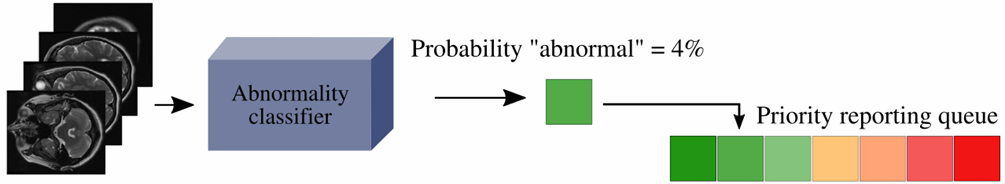
Figure: The trained classifier can be used to suggest the order in which head MRI examinations are reported by inserting images into a dynamic reporting queue based either on the predicted likelihood of showing an abnormal brain or both on the predicted category (normal vs abnormal) and the time spent in the queue.
Further Information
Wood, D. A.et al. (2022), "Deep learning models for triaging hospital head MRI examinations", Medical Image Analysis, 78, 102391, doi:10.1016/j.media.2022.102391.
Wood, D. A.et al. (2022), "Accurate brain‐age models for routine clinical MRI examinations", NeuroImage, 249, 118871, doi:10.1016/j.neuroimage.2022.118871.
Wood, D. A.et al. (2021), "Deep learning to automate the labelling of head MRI datasets for computer vision applications", European Radiology 32, 725–736, doi:10.1007/s00330-021-08132-0.